Paul Conduit The microtubule recipe for creating a wide variety of neuron shapes
CNRS researcher, Head of the “Microtubule regulation in multi-cellular animals” team at Institut Jacques Monod, Paris
- 2020 • ATIP-Avenir Program
- 2023 • Impulscience
Microtubules, which help make up the skeleton of cells, are required for neuronal growth and stability and help dictate the shape of these cells. By studying the elements necessary for microtubule formation, Paul Conduit and his team wish to know how controlling microtubule formation helps shape different neuronal types and resist neuronal degradation in the event of cellular stress.
Neurons come in a wide variety of shapes
Microtubules are protein-based structures that assemble into complex networks, specific to the shape and function of a cell. Most neurons form extensions called neurites, which enable them to receive and transmit information, and which can vary in length and complexity. Microtubules extend through these neurites, providing mechanical forces for their growth and stability and tracks for the transport of material throughout the neuron. Paul Conduit is particularly interested in the molecular mechanism which allows the formation of microtubules and wants to know if differentially regulating this process can contribute to variations in neuron shape.
Nucleation: where it all begins
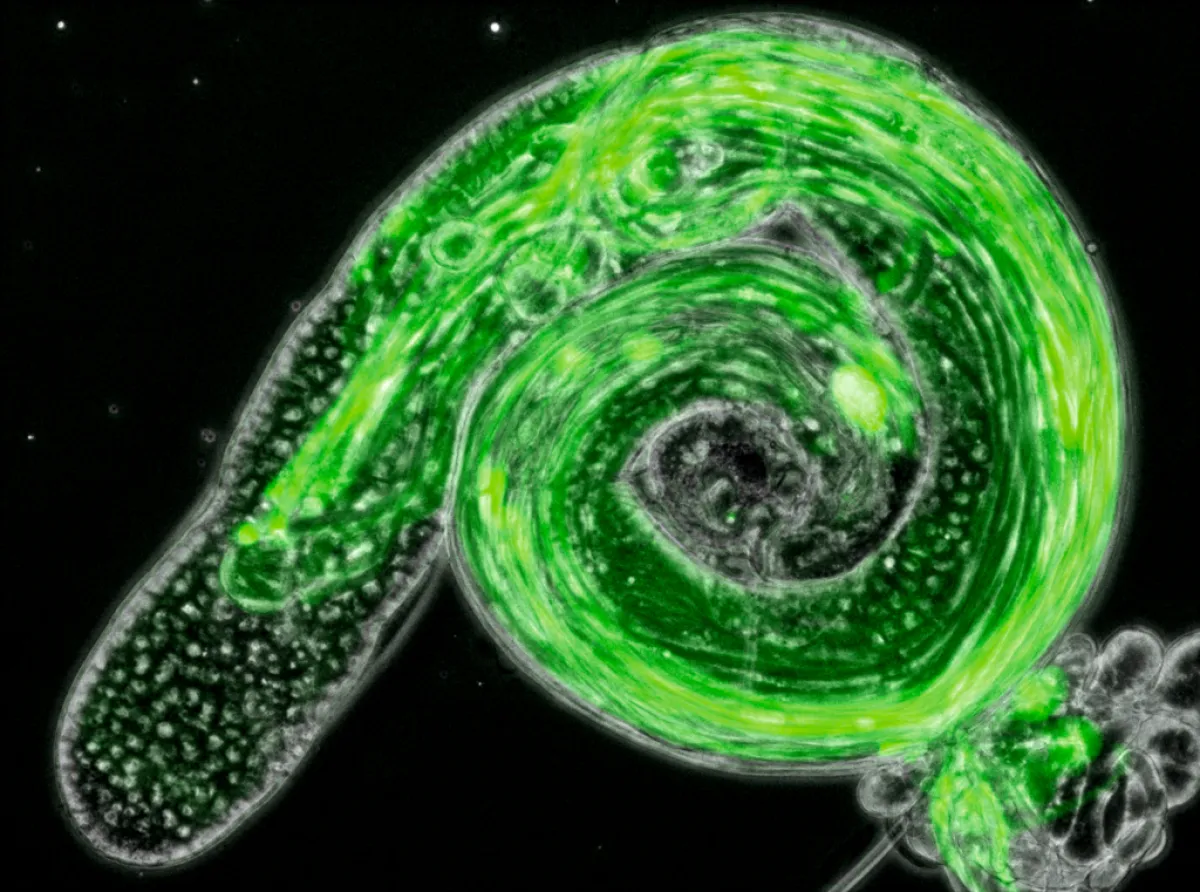
Microtubule formation doesn’t just happen randomly, it needs to be controlled in space and time. For this, groups of proteins called γ-tubulin ring complexes (γ-TuRCs) are recruited to specific locations in the cell where they trigger microtubule formation, known as nucleation. In his laboratory at the Institut Jacques Monod in Paris, Paul Conduit is interested in how this process is regulated so that cells can form their intricate microtubule networks. How does the regulation of γ-TuRCs vary in different types of neurons, during animal development and in response to cellular stress? These questions can be addressed using an elegant system: a family of neurons innervating the skin of the Drosophila fly whose highly varied shape is specific to their function. The advantage of these neurons is that they can be genetically manipulated, and that they can be easily visualized in the living fly using a microscope.
-
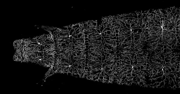
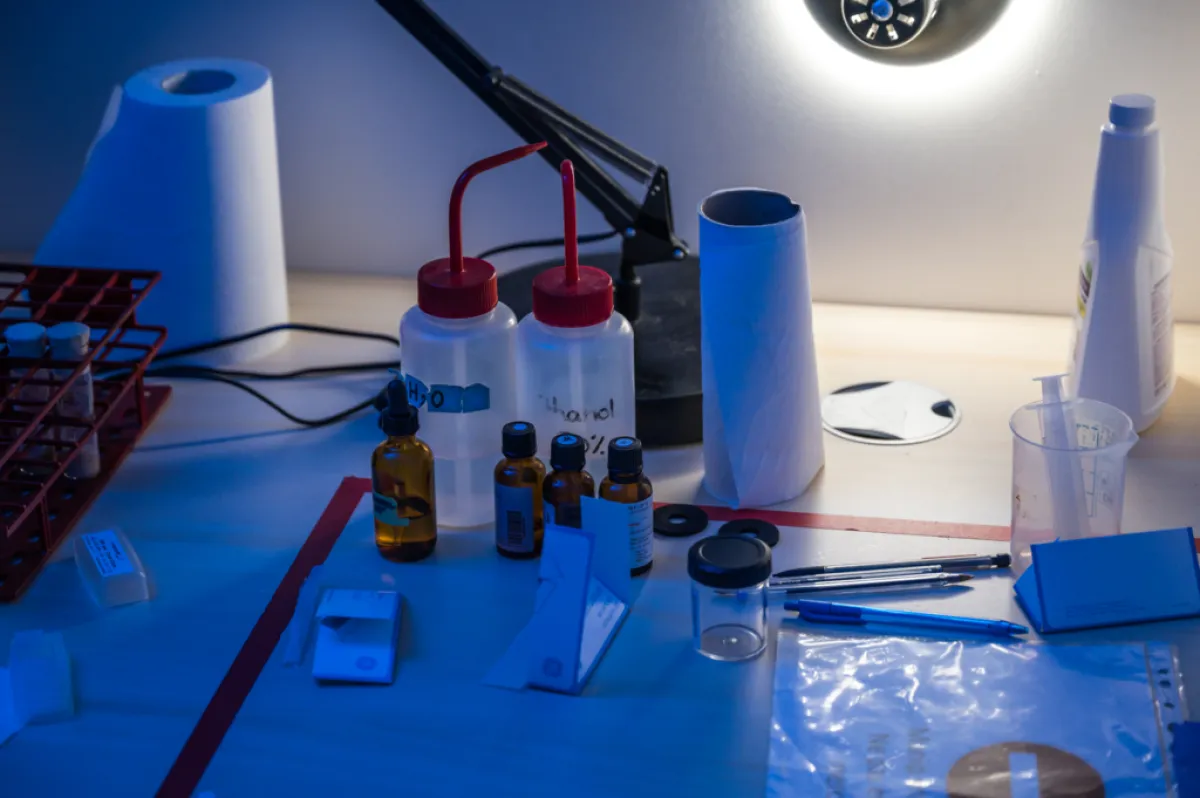
This image comes from the research work of Paul Conduit's team.
© Conduit Lab / Institut Jacques Monod -
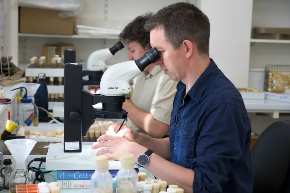
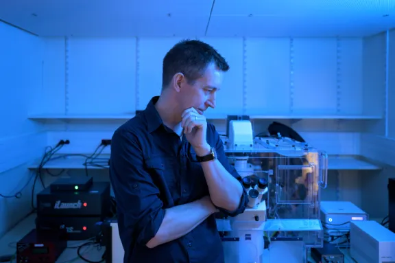
Paul Conduit in his laboratory, Paris, 2023.
© Alexandre Darmon/Art in Research pour la Fondation Bettencourt Schueller -
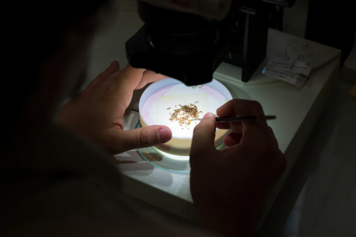
Paul Conduit's laboratory, Paris, 2023.
© Alexandre Darmon/Art in Research pour la Fondation Bettencourt Schueller -
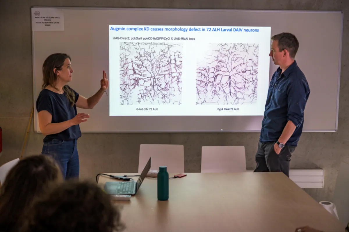
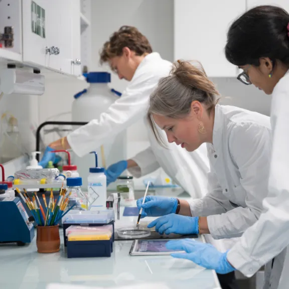
Paul Conduit and a member of his team, Paris, 2023.
© Alexandre Darmon/Art in Research pour la Fondation Bettencourt Schueller -
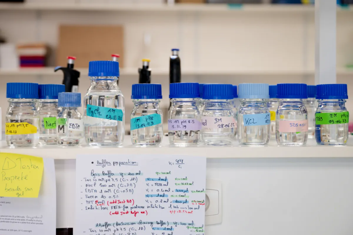
Paul Conduit's laboratory, Paris, 2023.
© Alexandre Darmon/Art in Research pour la Fondation Bettencourt Schueller -
Paul Conduit's laboratory, Paris, 2023.
© Alexandre Darmon/Art in Research pour la Fondation Bettencourt Schueller
Follow nucleation to understand the diversity of neuron shapes
Paul Conduit's team has developed methods to visualize γ-TuRCs and follow nucleation in Drosophila neurons in real time. This will allow them to reveal how this phenomenon is regulated in different types of neurons throughout development. Do nucleation sites form in the same place in highly branched neurons and in poorly branched neurons? Do γ-TuRCs always act with the same partners? Are γ-TuRCs required in adult neurons, once the microtubule networks have been established ?
In the case of stress, such as the sectioning of one neurite, nucleation increases to protect the neuron on a γ-TuRCs-dependent manner. But how are γ-TuRCs regulated to mediate this response? Is it possible to use them to protect neurons from degeneration?
This project supported by Impulscience® therefore aims to reveal new concepts on how neurons regulate their development, concepts which can be transposed to other types of cells and other organisms. In addition, the results obtained could help reveal ways to resist neurodegeneration during stress or suggest prospects for using γ-TuRCs as a target for anticancer treatments, which could be less toxic for neurons than current drugs targeting microtubules.
Paul Conduit in a few words
After studying biology at the University of Birmingham (United Kingdom), Paul Conduit devoted himself to the study of microtubule organization from centrosomes, which play an essential role in cell division. He first joined the laboratory of Doctor John Kilmartin at the Molecular Biology Laboratory of the Medical Research Council in Cambridge. He then completed his thesis on the formation of centrosomes with Jordan Raff at the Gurdon Institute in Cambridge.
During his postdoctoral stay at the University of Oxford, he closely studied the molecular mechanisms of centrosome formation. In 2015, he created his team in the department of zoology at the University of Cambridge where he began to study microtubule outside centrosomes, including in neurons. In 2020, Paul Conduit moved his team to the Institute Jacques Monod in Paris.

ATIP-Avenir Program
Since 2005, the Fondation Bettencourt Schueller has been a partner of the Inserm Avenir program. In 2009, the Avenir program merged with the ATIP program by the CNRS (National Center for Scientific Reasearch). The Foundation has since supported the ATIP-Avenir program which promotes the return or settlement in France of very high-level young researchers, with a research project of exceptional quality, and wishing to create their own team.
All the award-winnersImpulscience
Impulscience allocates 7 new grants each year to researchers in the life sciences. Focused on the mid-career, this program aims to support this crucial stage for the development of research projects.
All the award-winnersWorth reading

Simonetta Gribaldo

Ashley Nord
Anna Beyeler