Alexandre Baffet The fabulous fate of cerebral cortex stem cells
Alexandre Baffet, Inserm researcher, Head of the “Cell Biology of mammalian neurogenesis” team at the Institut Curie, Paris
- 2023 • Impulscience
The human cerebral cortex is known for its complexity and large size, which are key to the development of functions specific to humans, such as language. Alexandre Baffet aims to understand part of this unique complexity, that which comes from decisions made by neural stem cells to generate the amazing diversity of cells required for the cortex to function properly. The microscopy techniques he is developing with his research team will be invaluable allies in meeting the challenge.
The cells of the cerebral cortex are highly diverse
The cerebral cortex is the outermost part of the brain, and one the most complex. It is made up of a huge diversity of cell types, including several kinds of inhibitory and excitatory neurons as well as the glial cells (astrocytes and oligodendrocytes), which help the neurons carry out their functions. Most of these cells are generated by neural stem cells during embryonic development. Particularly plentiful in humans, they can divide to generate either neural stem cells, a particular type of neuron or a specific glial cell. The choices may vary between species as well as over time and depending on the cells’ location in the tissue (deeper or shallower in the cortex). However, how this diversity emerges during development and underpins differences in cell composition between different primates is not yet understood.
-
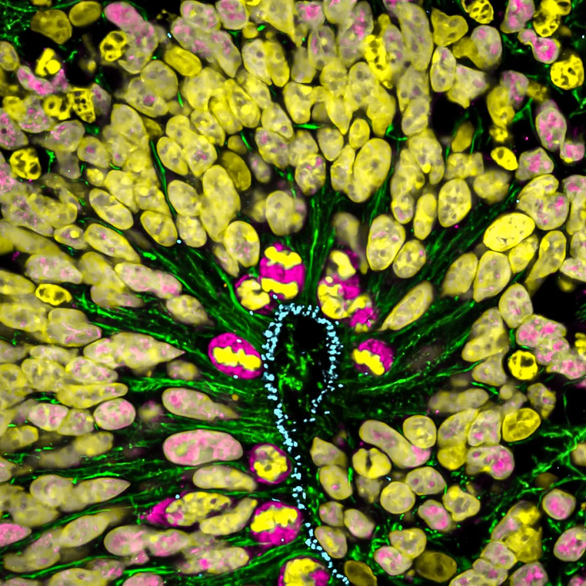
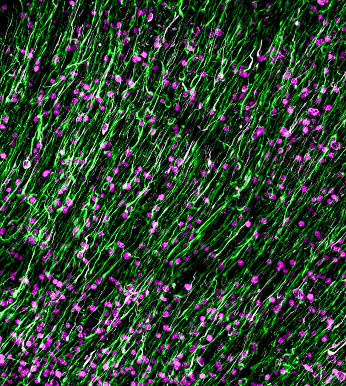
This image comes from the research work of Alexandre Baffet's team.
© Baffet Lab / Institut Curie -
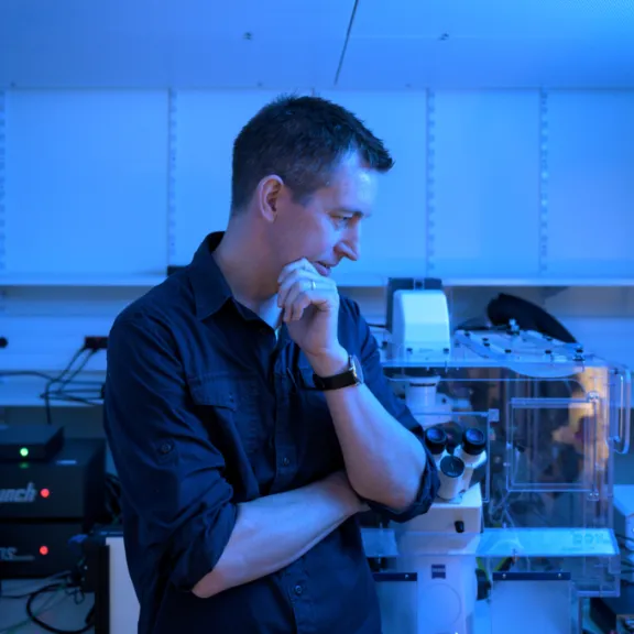
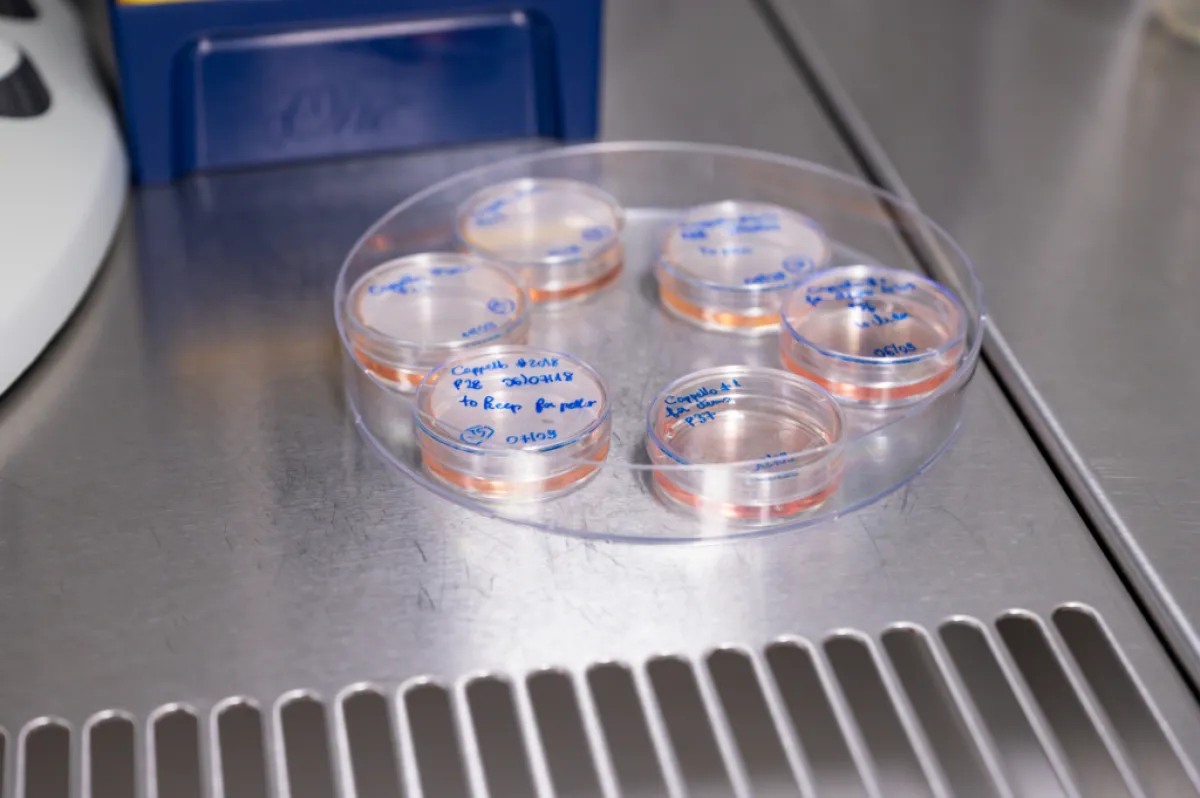
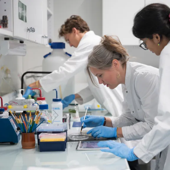
Alexandre Baffet's laboratory in Paris, 2023.
© Alexandre Darmon/Art in Research pour la Fondation Bettencourt Schueller -

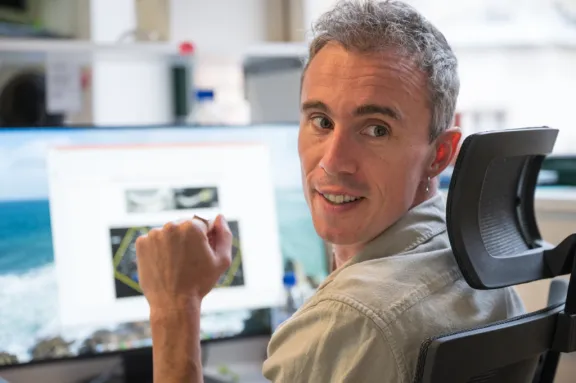
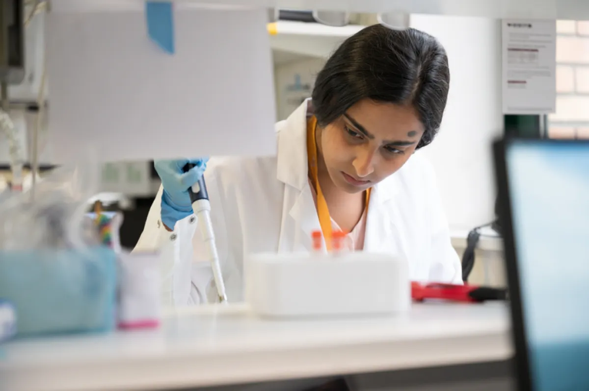
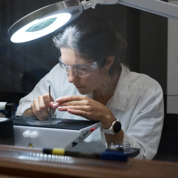
Alexandre Baffet's laboratory in Paris, 2023.
© Alexandre Darmon/Art in Research pour la Fondation Bettencourt Schueller -
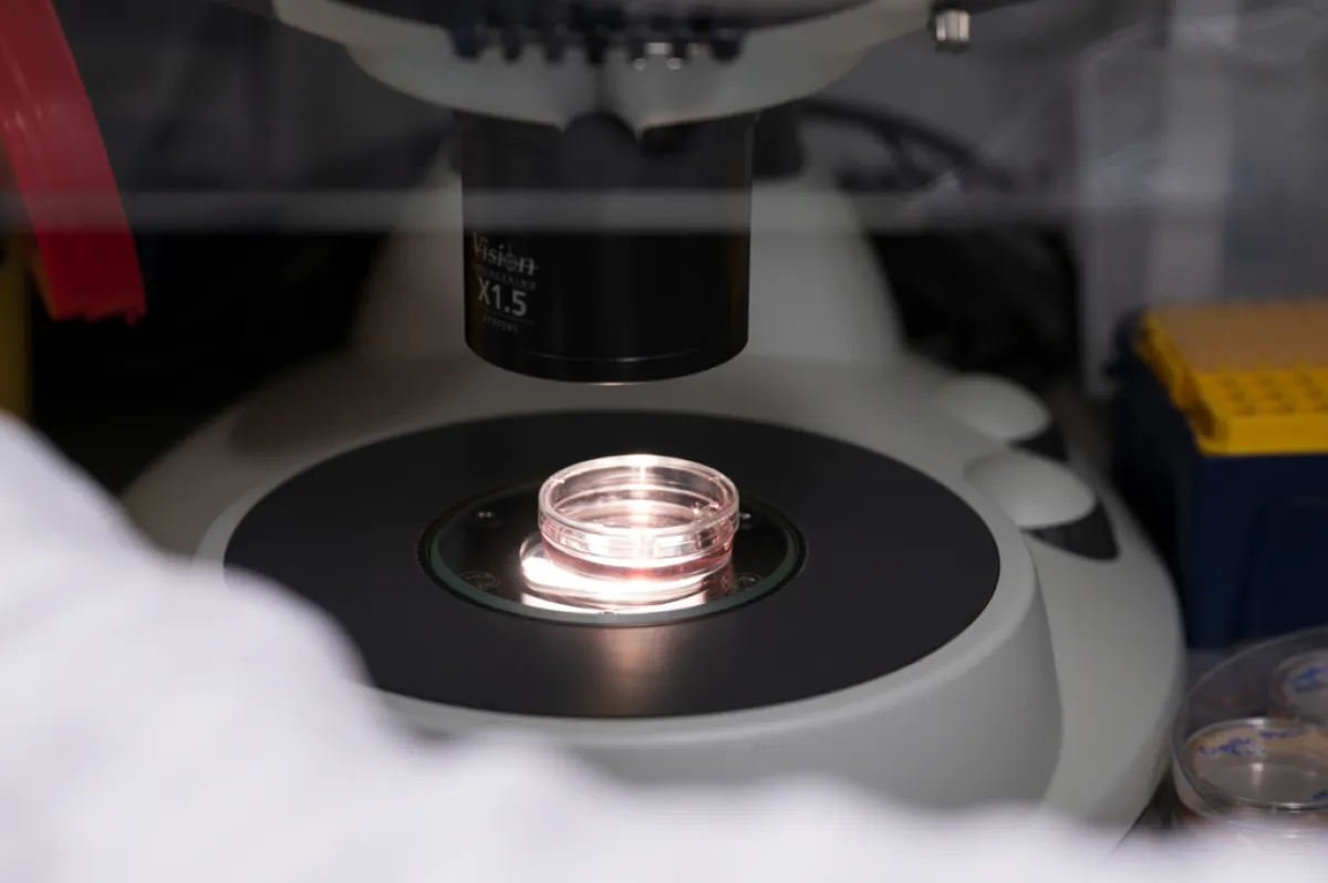
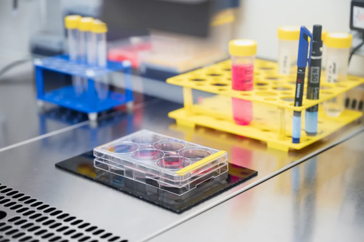
Alexandre Baffet's laboratory in Paris, 2023.
© Alexandre Darmon/Art in Research pour la Fondation Bettencourt Schueller -
Alexandre Baffet's laboratory in Paris, 2023.
© Alexandre Darmon/Art in Research pour la Fondation Bettencourt Schueller -
This image comes from the research work of Alexandre Baffet's team.
© Baffet Lab / Institut Curie -
Alexandre Baffet's laboratory in Paris, 2023.
© Alexandre Darmon/Art in Research pour la Fondation Bettencourt Schueller
Understanding the source of diversity
To understand how the choices made during the division of neural stem cells have impacted the complexity and evolution of the cerebral cortex of primates, it is necessary to be able to unravel the nature of these choices, identifying them in time and space.
Alexandre Baffet and his team at the Institut Curie plan to map neural stem cell division profiles using real-time imaging methods. They will use their recently developed technology to film neural stem cells while dividing within the tissue for several days and then identify the cell types produced. They will try to understand how the environment in which the cells evolve affects their fate and how this impact the variations observed within the primate order.
Models to answer evolution's big questions
The team uses so-called organoid models, which recreate certain features of the human cerebral cortex in vitro. The Impulscience® grant will allow them to develop advanced imaging methods on these tissues with the aim of accurately measuring the decisions neural stem cells make and understanding how their environment influences them. Furthermore, they will be able to identify the human-specific features underlying the cellular composition and size of our cerebral cortex by comparing them with those they will observe in organoids from chimpanzees, the species closest to humans.
Alexandre Baffet also wants to promote the technology used in his laboratory so that other research teams can use it. In this context, he is developing approaches to understand how derailed neural stem cell fate decisions can lead to paediatric brain tumours.
Alexandre Baffet in a few words
Live imaging to understand the dynamics of tissue formation is at the heart of Alexandre Baffet's career. He has developed this expertise since completing his PhD in 2010 at the Institut Jaques Monod and Université Paris VI, where he studied the transport of molecules in the fruit fly’s ovarian chamber. During his post-doctoral work at Columbia University in New York, he became interested in brain development and, again using live imaging, studied the migration of neural stem cells in the cortex of rodents. Since setting up his research team at the Institut Curie in 2016, he has been pushing the boundaries of live brain tissue imaging to study the development of the cortex.

Impulscience
Impulscience allocates 7 new grants each year to researchers in the life sciences. Focused on the mid-career, this program aims to support this crucial stage for the development of research projects.
All the award-winnersWorth reading
Anna Beyeler

Ashley Nord
Rebekka Wild