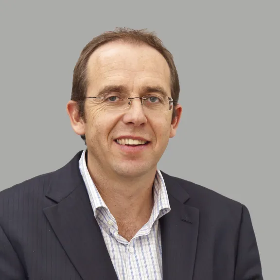
Thomas Lecuit Understanding the mechanics of cell building
Thomas Lecuit, Director of 1st grade research at the CNRS, Marseille, Director of the International Laboratory associated with the CNRS and the National Center for Biological Sciences, Bangalore, India
- 2015 • Liliane Bettencourt Prize for Life Sciences
How does a clump of cells become an embryo in a mother’s womb? Thomas Lecuit wants to answer this fascinating question. For years, he has been exploring the architecture and plasticity of epithelial tissues, which are made of juxtaposed cells lining the inner surface of organs.
How is an embryo made?
Our understanding of how embryos develop has made tremendous strides since the 1980s. It can even be summed up quite simply. In a way, genetics is the body’s "building plan". The genetic code is deployed in the form of chemical molecules that send instructions to the cells to tell them what they should do and what form they should take. This leads to the growth of cells that eventually form an embryo, an organ, etc.
Most discoveries in this area involve signaling pathways, genetic levers. Nothing, or almost nothing, allowed us to make a connection between biochemical and molecular information and mechanical changes in cells and tissues. So this is what Thomas Lecuit and his team focused on.
The three-dimensional formation of an organ
The researcher heralded a revolution by demonstrating in a 2004 Nature article that a motor protein takes apart the connections between epithelial cells in a polarized plane. This allows the intercalation of new cells necessary for tissue growth and the three-dimensional formation of the organ.
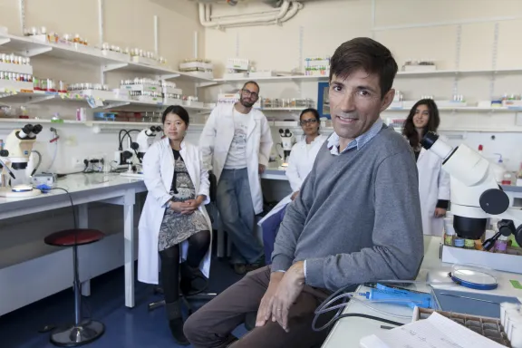
Dr. Lecuit and his multidisciplinary team study force regulation at the tissue level and 3D morphogenesis. He is also trying to answer a question that the scientific community has been asking for decades: how do the characteristic shapes of embryos and organs arise from changes in the shape of the cells themselves and from the regulation of contacts from one cell to another?
The foundation’s support
The support and funding provided by The Bettencourt Schueller Foundation allowed Dr. Lecuit to hire more team members to advance his research. The researchers aim to create a unified, quantitative model of morphogenesis that explains the different scales of organization and function allowing tissues to invaginate and elongate.
The laboratory uses the fruit fly as a model organism since its cell types and molecular mechanisms are similar to those of vertebrates. The research will shed light on other morphogenetic processes, particularly in epithelial organs such as the adult intestine and in the formation of cancerous tumors.
Thomas Lecuit in a few words
As a child, Thomas Lecuit was fascinated by butterflies. As a student at the École Normale Supérieure, he rediscovered the joys of naturalist observation during an internship on the embryogenesis of the fruit fly at Rockefeller University in New York. His Ph.D. research demonstrated that growth factors organize the adult fruit fly’s tissues along a gradient. Today this is taught in all biology classes.
As a post-doctoral fellow with Eric Wieschaus, he won the 1995 Nobel Prize for his work on the genetic control of early embryo development. He combines the exploration of fundamental mechanisms of morphogenesis with the study of physical forces and associated chemical events. His findings help to understand how a cell moves, finds its polarity in an epithelium, makes physical contacts and then modifies them to contribute to concerted morphogenetic movements.
Liliane Bettencourt Prize for Life Sciences
The Liliane Bettencourt Prize for Life Sciences rewards each year a researcher under the age of 45 for the excellence of their work and their remarkable contribution to their field of scientific research. This prize is awarded, depending on the year, to a researcher based in France or working in another European country. Thirty winners have been awarded since 1997. From 2023, prize rewards the laureate up to 100,000 euros.
All the award-winnersWorth reading
Scott Waddell
Monsef Benkirane
Cédric Blanpain